Background: Ventricular tachycardia is not uncommon after cardiac surgery, especially when non-sustained. However, given the potential for hemodynamic compromise, it warrants careful evaluation. In contrast to a VT with consistent ventricular morphology, Polymorphic Ventricular tachycardia is characterized by different morphologies of a wide QRS complex tachycardia. It is often poorly tolerated and should be assessed and treated immediately.
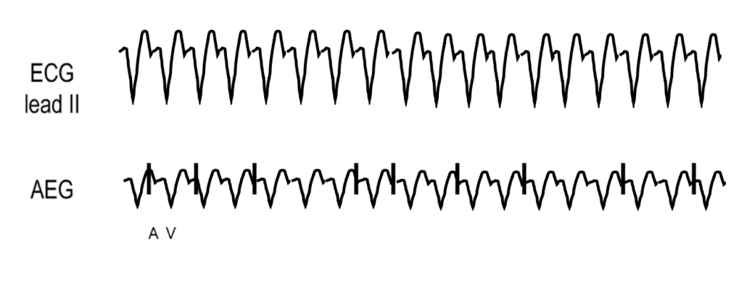
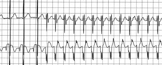
AEG: Ventricular tachycardia reveals a regular, ventricular rhythm with a wide QRS complex distinct from the baseline QRS appearance. The atrial to ventricular ratio is dependent on the AV nodal conduction, as VT can show both a 1:1 atrial to ventricular ratio, or more ventricular than atrial signals. In comparison to JET, VT has a morphology that is wide and distinct from the patient’s baseline QRS appearance. Here we show an idealized example of VT without retrograde conduction, and two examples of sinus rhythm followed by VT in post operative patients. Notice in both the patient examples, the atrial signals are ahead of the QRS during sinus, and are following the wide QRS complex on the AEG after VT onset.
Adenosine: Though there are exceptions, VT is generally not responsive to adenosine.
Atrial Overdrive Pacing: Again, though there are exceptions, VT is often not responsive to atrial overdrive pacing.