This simple atrial electrogram algorithm allows for the evaluation of the atrial and ventricular signals, their relative rates and their relationship to one another. This information allows accurate rhythm identification in the majority of patients. AEG can be used to distinguish both bradyarrhythmia and tachycardia but we will focus on the evaluation of tachyarrhythmias only.
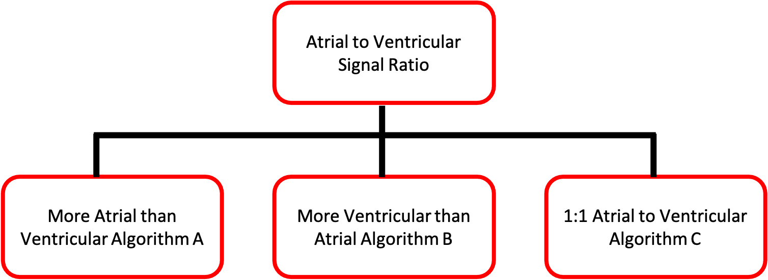
The first step is to determine the ratio of atrial to ventricular signals. Tachyarrhythmia can be classified by which chamber is “driving” the rate. If there are more atrial signals than ventricular signals, the tachyarrhythmia is an atrial driven such as atrial fibrillation, intra-atrial reentrant tachycardia (atrial flutter) atrial ectopic tachycardia (AET) or sinus tachycardia. Similarly, if there are more ventricular signals then atrial signals, the rhythm is ventricular driven such as with ventricular tachycardia or junctional ectopic tachycardia. Rhythms which have a 1:1 atrial:ventricular ratio can be more challenging to diagnose as they can be either atrial or ventricular in origin.
After presentation of the algorithm for tachyarrhythmia diagnosis, a more complete discussion of the AEG for each arrhythmia will follow.
Many arrhythmias can also be classified based on their response to Adenosine and/or atrial overdrive pacing. To help further characterize the type of arrhythmia, you can follow this link to lean how different arrhythmias react to adenosine or pacing. This is also clarified when discussing each individual arrhythmia.